

lv filling pressure | elevated left sided filling pressures
$283.00
In stock
Left ventricular filling pressure (LVFP) is a critical determinant of cardiac performance, reflecting the pressure within the left ventricle during diastole, the relaxation phase of the heart cycle. It represents the preload, the degree of stretch on the ventricular muscle fibers at the end of diastole, just before contraction. This preload, dictated primarily by LVFP, profoundly influences stroke volume, the amount of blood ejected with each heartbeat, through the Frank-Starling mechanism. Understanding LVFP, its normal range, factors affecting it, and the implications of elevated levels, particularly in heart failure (HF), is crucial for effective diagnosis and management of cardiovascular disease.
The Significance of LV Filling Pressure
The heart's primary function is to pump blood efficiently throughout the body, delivering oxygen and nutrients to tissues and removing waste products. The Frank-Starling mechanism elegantly describes the relationship between preload (LVFP) and stroke volume. In essence, within physiological limits, the more the heart is stretched during filling, the more forcefully it will contract, and the greater the stroke volume. This ensures that the heart adapts to varying demands placed upon it, such as during exercise or periods of stress.
LVFP is not a static value. It is influenced by several factors including:
* Blood Volume: The amount of circulating blood directly affects the volume returning to the heart (venous return), and consequently, the pressure required to fill the left ventricle.
* Venous Tone: The constriction or dilation of veins influences the blood volume returning to the heart.
* Atrial Contraction: The forceful contraction of the left atrium contributes to the final filling of the left ventricle, slightly increasing LVFP.
* Ventricular Compliance: This refers to the ability of the left ventricle to relax and expand during diastole. A stiff, non-compliant ventricle requires higher pressures to achieve adequate filling.
* Heart Rate: Rapid heart rates shorten diastolic filling time, potentially affecting LVFP and stroke volume.lv filling pressure
* Valve Function: Mitral valve stenosis (narrowing) or regurgitation (leakage) can significantly alter LVFP.
* Pericardial Pressure: Pressure from the pericardial sac surrounding the heart can impede ventricular filling.
LV Filling Pressure Normal Range
Determining the normal range for LVFP is complex and depends on the method of measurement. Invasive methods, such as right heart catheterization, provide the most accurate assessment. Non-invasive methods, like echocardiography, can estimate LVFP but are less precise.
Generally, the normal LV end-diastolic pressure (LVEDP), a key indicator of LVFP, is considered to be between 5-12 mmHg. However, this range can vary slightly depending on the laboratory and the individual patient.
It is essential to remember that a single measurement of LVFP is just a snapshot in time. Clinicians often look at trends and consider the patient's overall clinical picture when interpreting LVFP values. Factors like age, body size, and the presence of other medical conditions can influence what is considered "normal" for a particular individual.
Methods of Assessing LV Filling Pressure
Several methods are employed to assess LVFP, each with its own advantages and limitations:
* Right Heart Catheterization (RHC): This invasive procedure involves inserting a catheter into a vein, typically in the neck or groin, and threading it through the right side of the heart into the pulmonary artery. RHC allows for direct measurement of pressures in the right atrium, right ventricle, pulmonary artery, and pulmonary capillary wedge pressure (PCWP). PCWP is often used as an estimate of LVFP, although it is not a direct measurement and can be affected by pulmonary vascular disease. RHC is considered the gold standard for assessing LVFP but is reserved for more complex cases or when non-invasive methods are inconclusive.
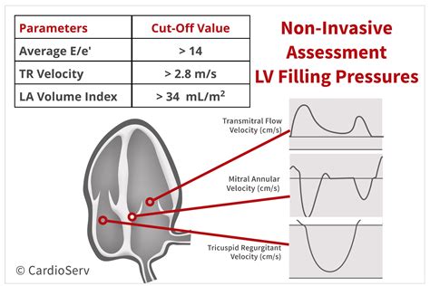
* Echocardiography: This non-invasive imaging technique uses ultrasound waves to create images of the heart. Echocardiography can assess various parameters related to LV filling, including:
* E/A Ratio: This ratio compares the peak early diastolic filling velocity (E wave) to the peak late diastolic filling velocity due to atrial contraction (A wave). A normal E/A ratio suggests adequate LV relaxation and compliance.
* E/e' Ratio: This ratio divides the E wave velocity by the early diastolic mitral annular velocity (e'). The e' velocity reflects the relaxation of the left ventricle. An elevated E/e' ratio is a strong indicator of elevated LVFP.
* Left Atrial Size: A dilated left atrium can be a sign of chronically elevated LVFP.
* Pulmonary Vein Flow Patterns: Abnormal pulmonary vein flow patterns can also suggest elevated LVFP.
* Tricuspid Regurgitation Velocity (TRV): TRV can be used to estimate pulmonary artery systolic pressure (PASP). Elevated PASP can be a consequence of chronically elevated LVFP.
* Cardiac Magnetic Resonance Imaging (MRI): Cardiac MRI provides detailed images of the heart's structure and function. While not routinely used to directly measure LVFP, MRI can assess ventricular volumes, ejection fraction, and wall motion abnormalities, providing valuable information about overall cardiac function and potential causes of elevated LVFP.
Elevated LV Filling Pressure: A Hallmark of Heart Failure
Additional information
| Dimensions | 9.6 × 4.2 × 1.2 in |
|---|