lv meaning medical | what is Lv systolic dysfunction
$189.00
In stock
In the realm of medicine, abbreviations and acronyms are frequently used to streamline communication and documentation. One such abbreviation, "LV," commonly encountered in cardiology, stands for Left Ventricle. Understanding the significance of LV and the conditions associated with it is crucial for comprehending various cardiac ailments. This article delves into the meaning of LV in medical terms, focusing primarily on Left Ventricular Hypertrophy (LVH) and other related conditions like left ventricular hypokinesis, impairment, and systolic dysfunction, exploring their causes, dangers, and implications for overall health.
LV in Medical Terms: The Left Ventricle Explainedlv meaning medical
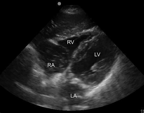
The heart, a vital organ responsible for circulating blood throughout the body, is composed of four chambers: two atria (upper chambers) and two ventricles (lower chambers). The left ventricle (LV) is the heart's main pumping chamber. It receives oxygenated blood from the left atrium and pumps it into the aorta, the body's largest artery, which then distributes the blood to the rest of the body. Due to the immense pressure required to pump blood throughout the systemic circulation, the left ventricle possesses the thickest walls of all the heart chambers.
Therefore, any abnormality affecting the structure or function of the left ventricle can have profound consequences on overall cardiovascular health. LV is a cornerstone term in cardiology, appearing in diagnoses, test results (like echocardiograms and EKGs), and treatment plans.
Left Ventricular Hypertrophy (LVH): Thickening of the Heart Wall
Left Ventricular Hypertrophy (LVH) refers to the thickening of the walls of the left ventricle. This thickening isn't inherently beneficial; instead, it's often a sign that the heart is working harder than it should to pump blood. The increased workload can lead to various complications.
* How LVH Develops: LVH typically develops as a response to increased pressure or volume overload on the left ventricle. The heart muscle cells (cardiomyocytes) enlarge to compensate for the increased workload. Over time, this enlargement can lead to structural changes in the heart wall.
* Consequences of LVH:
* Stiffening of the Heart Wall: The thickened heart wall can become stiff, making it harder for the left ventricle to relax and fill properly with blood during diastole (the relaxation phase of the heart). This reduced filling capacity can lead to decreased cardiac output, meaning the heart pumps less blood with each beat.
* Increased Risk of Arrhythmias: LVH can disrupt the heart's electrical system, increasing the risk of arrhythmias (irregular heartbeats), some of which can be life-threatening.
* Increased Risk of Heart Failure: Over time, LVH can weaken the heart muscle, eventually leading to heart failure, a condition in which the heart is unable to pump enough blood to meet the body's needs.
* Increased Risk of Sudden Cardiac Death: LVH is a known risk factor for sudden cardiac death, especially in individuals with underlying heart conditions.
* Increased Myocardial Oxygen Demand: The enlarged heart muscle requires more oxygen to function. If the blood supply to the heart is insufficient (e.g., due to coronary artery disease), it can lead to angina (chest pain) and myocardial ischemia (reduced blood flow to the heart muscle).
* Causes of LVH:
* High Blood Pressure (Hypertension): Chronic hypertension is the most common cause of LVH. The left ventricle has to work harder to pump blood against the elevated pressure in the arteries.
* Aortic Stenosis: Aortic stenosis is a narrowing of the aortic valve, which restricts blood flow from the left ventricle into the aorta. This forces the left ventricle to pump harder to overcome the obstruction.
* Hypertrophic Cardiomyopathy (HCM): HCM is a genetic condition that causes abnormal thickening of the heart muscle, often affecting the left ventricle.
* Mitral Regurgitation: Mitral regurgitation is a condition in which the mitral valve, which separates the left atrium and left ventricle, doesn't close properly, allowing blood to leak back into the left atrium during ventricular contraction. This increases the workload on the left ventricle.
* Aortic Regurgitation: Aortic regurgitation is when the aortic valve doesn't close properly, allowing blood to leak back into the left ventricle after it has been pumped into the aorta.
* Obesity: Obesity is associated with increased blood volume and cardiac output, which can lead to LVH.
* Sleep Apnea: Sleep apnea can cause intermittent hypoxia (low oxygen levels) and increased blood pressure, both of which can contribute to LVH.
* Endurance Athletes: In some cases, intense endurance training can lead to physiological LVH, which is considered a normal adaptation to exercise. However, it's important to differentiate physiological LVH from pathological LVH.
* Diagnosis of LVH:
* Electrocardiogram (ECG or EKG): An ECG can detect electrical patterns indicative of LVH.
* Echocardiogram: An echocardiogram (ultrasound of the heart) is the most common and reliable method for diagnosing LVH. It can measure the thickness of the left ventricular wall and assess the heart's overall structure and function.
Additional information
| Dimensions | 5.7 × 5.1 × 3.8 in |
|---|